
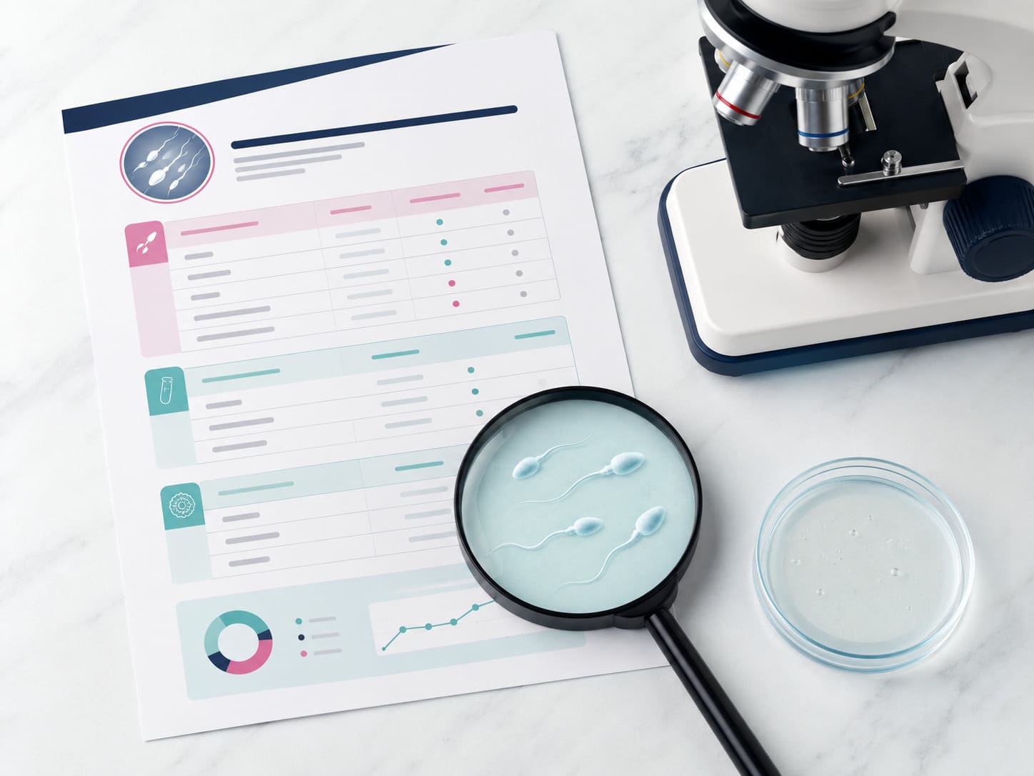
Most men receive their semen analysis report as a printout of numbers — without anyone explaining what those numbers mean. A column of parameters, reference ranges, and a result of ‘abnormal’ or ‘borderline normal.’ For many couples, this report is the first piece of clinical evidence in their fertility journey — and the confusion or fear it generates is unnecessary.
A semen analysis is not a simple pass-or-fail test. Each parameter tells a different part of the story about sperm health, and a result that looks alarming in isolation often looks very different in clinical context. More importantly, knowing what your report means — specifically — is the difference between panic and informed decision-making.
In this guide — written from clinical practice at Wellspring IVF & Women’s Hospital, Ahmedabad — I will walk through every parameter on a standard semen analysis report: what it measures, what the current WHO 2021 (6th Edition) reference range is, what an abnormal result means clinically, and — critically — what treatment options exist when a parameter falls below the threshold. By the time you finish this guide, you will be able to read your own report with clarity.
What Is a Semen Analysis? Purpose and Limitations
A semen analysis (SA) is the primary diagnostic test for male fertility — assessing the quantity, movement, and structure of sperm in an ejaculate sample. It is the clinical starting point for evaluating the male contribution to a couple’s fertility, and it is typically the first investigation recommended when a couple has been trying to conceive for 12 months without success (or 6 months if the female partner is over 35).
However, understanding its limitations is as important as understanding its results:
- It is not a pregnancy test: A normal semen analysis does not guarantee fertility. Fertilisation involves complex sperm-egg interactions that a standard SA cannot assess.
- Significant intra-individual variability: A single SA result can be influenced by illness in the preceding 3 months (the full sperm maturation cycle — spermatogenesis — takes approximately 74 days), stress, recent fever, alcohol consumption, or sexual abstinence duration. A single abnormal result should always be confirmed with a repeat test after 6–12 weeks.
- Standard SA does not assess sperm DNA: DNA Fragmentation Index (DFI) — one of the most clinically significant predictors of IVF outcome — requires a separate, specific test not included in routine semen analysis. This is covered in detail below.
- Normal SA does not rule out male factor infertility: Approximately 15% of men with unexplained infertility have a normal semen analysis but high DNA fragmentation. A ‘normal’ report from a standard SA should not be the end of investigation when IVF failure or recurrent miscarriage is the clinical picture.
Ideal sample collection: Collected after 2–5 days of sexual abstinence. Produced by masturbation into a sterile container. Delivered to the laboratory within 60 minutes of collection, kept at body temperature. Labs at Wellspring IVF have an on-site andrology laboratory for immediate analysis, minimising handling variability.
The WHO 2021 (6th Edition) Reference Ranges — The Updated Standard
In 2021, the World Health Organisation published the 6th Edition of the Laboratory Manual for Examination and Processing of Human Semen — the current international gold standard. The 6th Edition updated reference limits from the previous 2010 (5th Edition) values. If your report shows reference ranges from an older laboratory using the 2010 criteria, your results may be flagged differently. Always confirm which edition your laboratory is using.
The table below shows all key parameters with current WHO 2021 lower reference limits (5th percentile of fertile men):
| Parameter | WHO 2021 Lower Reference Limit | Previous WHO 2010 Limit | Clinical Significance |
|---|---|---|---|
| Semen Volume | ≥1.4 mL | ≥1.5 mL | Accessory gland function; very low volume can indicate ejaculatory duct obstruction |
| pH | ≥7.2 | ≥7.2 | Acidity may indicate infection or absent seminal vesicle secretions |
| Sperm Concentration | ≥16 million/mL | ≥15 million/mL | Primary measure of sperm production per unit volume |
| Total Sperm Count | ≥39 million/ejaculate | ≥39 million/ejaculate | Total sperm output — accounts for both concentration and volume |
| Total Motility (PR+NP) | ≥42% | ≥40% | Combined progressive + non-progressive movement |
| Progressive Motility | ≥30% | ≥32% | The clinically critical measure — sperm moving forward purposefully |
| Immotile Sperm | <58% | <60% | Any result with majority immotile sperm warrants vitality assessment |
| Morphology (Kruger) | ≥4% normal forms | ≥4% normal forms | Strictest assessment of sperm shape — most sensitive predictor of fertilisation capacity |
| Vitality (Live sperm) | ≥54% | ≥58% | Distinguishes immotile-but-alive from dead sperm (necrozoospermia) |
| Leukocytes | <1 million/mL | <1 million/mL | Above threshold indicates infection/inflammation requiring investigation |
Note: These are lower reference limits — the 5th percentile of men with proven fertility. They are not ‘optimal’ values. Fertility is possible at levels below these thresholds and not guaranteed above them. The clinical picture — including female partner factors and the couple’s full history — always determines treatment strategy.
Parameter 1: Semen Volume
What it measures: The total volume of fluid ejaculated. Semen is not sperm — sperm cells account for a tiny fraction of semen volume. The bulk of semen is produced by the seminal vesicles (approximately 65%), prostate gland (approximately 30%), and bulbourethral glands.
Normal range (WHO 2021): ≥1.4 mL
Low Volume (Hypospermia): < 1.4 mL
Low semen volume with normal sperm concentration can still mean an adequate total sperm count — the key is always the total count, not concentration alone. However, very low volume (under 1.0 mL) may indicate:
- Ejaculatory duct obstruction
- Retrograde ejaculation (sperm entering the bladder instead of being expelled)
- Bilateral absence of the vas deferens (CBAVD) — associated with CFTR gene mutations
- Androgen deficiency
- Incomplete collection of the sample
High Volume (Hyperspermia): > 6 mL
High volume can dilute sperm concentration, reducing total motile sperm count despite an apparently adequate sperm count reading. It may also indicate prostate gland hyper-secretion.
Parameter 2: pH
What it measures: Acidity or alkalinity of the ejaculate. Normal semen is slightly alkaline (pH ≥7.2) — this protects sperm from the acidic environment of the vagina.
Acidic pH (<7.0) combined with low volume may indicate obstruction or absence of the seminal vesicles. The prostate secretion is acidic; seminal vesicle secretion is alkaline. When seminal vesicle secretion is absent, the mixture becomes acidic. This pattern — low volume + acidic pH + azoospermia — strongly suggests obstructive cause and guides further investigation toward imaging.
Parameter 3: Liquefaction and Viscosity
Liquefaction: Semen initially coagulates on ejaculation (due to seminal vesicle proteins) and then liquefies within 15–30 minutes (due to prostate enzymes). A report noting ‘incomplete liquefaction’ at 60 minutes suggests prostate gland dysfunction and may impair sperm motility and migration.
Viscosity: Abnormally thick or viscous semen may indicate infection or gland dysfunction and can physically impair sperm movement. High viscosity is a separate finding from incomplete liquefaction — thick semen can still be fully liquefied. Both findings warrant clinical follow-up.
Parameter 4: Sperm Concentration and Total Sperm Count
These are the two most commonly misread parameters on a semen analysis — and the distinction between them is clinically critical.
Sperm Concentration vs Total Sperm Count
| Parameter | Definition | Why It Matters |
|---|---|---|
| Sperm Concentration | Number of sperm per millilitre (million/mL) | Reflects density but not the whole picture — a small-volume sample with high concentration may have low total count |
| Total Sperm Count | Concentration × Semen Volume = total sperm per ejaculate (millions) | The clinically more meaningful number — total sperm delivered per ejaculation |
Example: A report showing 20 million/mL concentration with 1.5 mL volume gives a total count of 30 million — which falls below the WHO 2021 reference limit of 39 million, even though the concentration alone appears borderline normal. Always check the total count, not just concentration.
Classification of Sperm Count Results
| Diagnosis | Definition | Clinical Implication |
|---|---|---|
| Normozoospermia | All parameters within WHO 2021 limits | No sperm count barrier to conception — other factors investigated |
| Oligozoospermia | Total count < 39 million or concentration < 16 million/mL | Mild: 10–39M/ejaculate | Moderate: 5–10M | Severe: < 5M | ICSI typically indicated |
| Cryptozoospermia | Extremely rare sperm (< 100,000/mL) — only found after centrifugation | Sperm may still be extractable; surgical retrieval often considered |
| Azoospermia | No sperm in ejaculate (confirmed on centrifuged pellet) | Requires further workup — obstructive vs non-obstructive — see below |
Azoospermia — zero sperm count — is the most feared finding, but it does not mean parenthood is impossible. Surgical sperm retrieval via TESE / PESA is successful in a significant proportion of cases. For a full explanation of what an azoospermia diagnosis means and what options exist, see our dedicated page: Azoospermia (Nil Sperm Count) — Treatment at Wellspring.
For low but present sperm count — oligozoospermia — ICSI (Intracytoplasmic Sperm Injection) allows fertilisation with very low sperm counts. See: Oligospermia — Causes, Grading & IVF Options.
Parameter 5: Sperm Motility — Total, Progressive, and Non-Progressive
Motility is arguably the most clinically important group of parameters on a semen analysis — because sperm that cannot move cannot reach and fertilise the egg.
Understanding the Three Motility Categories
| Motility Type | WHO Symbol | Definition | WHO 2021 Lower Limit |
|---|---|---|---|
| Progressive motility | PR | Sperm moving forward — either linearly or in large circles | ≥30% |
| Non-progressive motility | NP | Sperm moving but not progressing — tail movement without forward travel | No specific lower limit; adds to total motility |
| Immotile | IM | No movement observed | No specific lower limit; total PR+NP must be ≥42% |
| Total motility | PR+NP | Sum of progressive and non-progressive | ≥42% |
Clinical Diagnoses Based on Motility
- Asthenozoospermia: Total motility < 42% and/or progressive motility < 30%. The most common sperm parameter abnormality in Indian male fertility clinics. Causes include varicocele, genital tract infection, oxidative stress, lifestyle factors (smoking, heat exposure, obesity), and sperm DNA damage.
- Necrozoospermia: The majority of sperm are immotile AND non-viable (dead). Distinguished from asthenozoospermia — which can have many immotile-but-alive sperm — by the vitality test.
- Asthenoteratozoospermia: Combined motility and morphology defect — the most common combined SA abnormality.
For a complete clinical explanation of poor sperm motility, its causes, and treatment pathways, see: Poor Sperm Motility — Causes, Testing & Treatment at Wellspring.
Parameter 6: Sperm Morphology — Kruger Strict Criteria
Morphology — the shape and structural integrity of sperm — is the most misunderstood and most anxiety-provoking parameter for most patients. A report showing only 3–4% normal forms frequently triggers alarm. Here is the clinical context that changes how that number should be read:
What ‘Normal Morphology’ Actually Means
Under Kruger Strict Criteria — the most stringent assessment method and the one used by most fertility laboratories — a sperm is classified as ‘normal’ only if it meets exacting criteria for head shape, midpiece structure, and tail integrity. By this standard, even fertile men typically have only 4–15% morphologically normal sperm. The remaining 85–96% are structurally abnormal in fertile men.
Key Insight on Morphology:
Low morphology does not mean your sperm cannot fertilise an egg — it means a higher proportion of your sperm would have difficulty penetrating the egg unenassisted. With ICSI, where a single selected sperm is directly injected into the egg, morphology has significantly less impact on fertilisation outcome than it does in natural conception or IUI. Studies show ICSI fertilisation rates are comparable across a wide range of morphology results when an experienced embryologist selects the injection sperm.
Morphology Classification
| Result | Kruger % | Clinical Category | Recommended Approach |
|---|---|---|---|
| Normal | ≥4% | Normozoospermia (morphology) | No specific morphology barrier to IVF/ICSI |
| Borderline | 2–3% | Mild Teratozoospermia | IVF with ICSI standard recommendation; success rates unaffected with ICSI |
| Severe | 0–1% | Severe Teratozoospermia | ICSI with IMSI (high-magnification sperm selection) may be considered; DFI testing recommended |
| Globozoospermia | 0% — round-headed sperm | Rare morphological defect | ICSI may have lower rates; specialised calcium ionophore protocols may be needed |
For severe teratozoospermia, IMSI (Intracytoplasmic Morphologically-Selected Sperm Injection) — which uses 6,600× magnification to select the most structurally intact sperm — may improve embryo development outcomes in selected cases.
Parameter 7: Vitality (Live vs Dead Sperm)
Vitality measures what proportion of sperm are alive — regardless of whether they are moving. This parameter is specifically requested when immotility is high, to distinguish between:
- Asthenozoospermia (immotile but alive): Sperm are alive but cannot move. Causes include structural flagellar defects (e.g., Primary Ciliary Dyskinesia) or biochemical energy production failure. Vitality ≥54% (WHO 2021). ICSI can achieve fertilisation even with high asthenozoospermia since the sperm is mechanically injected.
- Necrozoospermia (dead sperm): Vitality significantly below 54% indicates that most immotile sperm are also dead. This is a different clinical problem with different causes — testicular failure, epididymal defects, or sperm storage dysfunction. Requires additional investigations including hormonal panel and potentially testicular biopsy.
Clinical note: When vitality is very low but concentration is normal, consider sperm DNA fragmentation testing — oxidative stress causing DNA damage is a common pathway that kills sperm before ejaculation.
Parameter 8: Leukocytes (White Blood Cells in Semen)
Normal semen contains a small number of white blood cells (leukocytes). The WHO 2021 upper limit is 1 million/mL. Above this threshold — termed leukocytospermia — the finding has two important implications:
- Infection / Inflammation: Leukocytospermia may indicate bacterial infection of the genital tract (prostatitis, epididymitis, seminal vesiculitis) or asymptomatic inflammation. A culture of the semen and urine should be requested. Treatment with appropriate antibiotics typically resolves the finding and may improve sperm parameters.
- Oxidative Stress and DNA Damage: Activated neutrophils produce reactive oxygen species (ROS) that damage sperm cell membranes and DNA. Even without overt infection, elevated leukocytes are associated with higher sperm DNA fragmentation. If leukocytospermia is persistent despite antibiotic treatment, DFI testing is strongly recommended.
Parameter 9: Sperm DNA Fragmentation — The Test Most Reports Miss
DNA Fragmentation Index (DFI) is the single most underutilised parameter in male fertility assessment in India — and the one most likely to explain unexplained IVF failure and recurrent miscarriage when the standard semen analysis appears normal.
What Is Sperm DNA Fragmentation?
Sperm DNA fragmentation refers to breaks or damage within the genetic material carried by the sperm cell. A certain degree of DNA damage is normal and repaired by the egg at fertilisation. However, when DFI exceeds a critical threshold — typically >25–30% by the SCSA (Sperm Chromatin Structure Assay) method — the egg’s repair capacity may be overwhelmed, leading to fertilisation failure, poor-quality embryos, or early miscarriage.
DFI is NOT routinely included in standard semen analysis. It requires a separate test — SCSA, TUNEL, or SCD (Sperm Chromatin Dispersion) — specifically requested by the clinician.
DFI Reference Ranges
| DFI Level | Clinical Interpretation | Recommended Action |
|---|---|---|
| < 15% | Normal — low fragmentation | No specific DFI-related intervention required |
| 15–25% | Borderline — elevated fragmentation | Lifestyle optimisation; antioxidant supplementation; repeat in 3 months |
| 25–30% | High fragmentation — clinical significance likely | ICSI with surgical sperm (TESE) may outperform ejaculated sperm; IMSI consideration |
| > 30% | Very high — directly implicated in IVF failure | Surgical sperm retrieval from testicular source; DFI reduction protocol |
The most effective intervention for high DFI is testicular sperm extraction (TESE), because testicular sperm — extracted directly from the site of production — consistently have lower DFI than ejaculated sperm. ICSI with testicular sperm in high-DFI cases has demonstrated significantly improved embryo quality and reduced miscarriage rates in multiple clinical studies.
For a detailed explanation of DFI, its causes, how it is tested, and how Wellspring addresses it clinically: Sperm DNA Fragmentation — DFI Testing, Causes & Treatment.
Understanding Combined Diagnoses: OAT Syndrome and Other Patterns
When multiple parameters are abnormal simultaneously, the combined finding has a specific clinical diagnosis:
| Combined Diagnosis | Parameters Affected | Clinical Significance |
|---|---|---|
| OAT Syndrome | Oligozoospermia + Asthenozoospermia + Teratozoospermia | The most common combined male infertility diagnosis. IVF with ICSI is the standard treatment pathway. |
| Asthenoteratozoospermia | Low motility + Low morphology (count normal) | ICSI with IMSI consideration if morphology is severe |
| Oligoasthenozoospermia | Low count + Low motility | ICSI; evaluate for varicocele and testosterone/LH/FSH panel |
| Total Asthenozoospermia | 0% motility, all parameters otherwise normal | Vitality test mandatory; if vital — PCD or axonemal defect workup |
| Azoospermia | No sperm in ejaculate | Hormonal panel + TRUS + testicular biopsy to classify obstructive vs non-obstructive |
What to Do After Reading Your Report: Clinical Next Steps by Result
Your Report Is Normal — What Next?
A normal semen analysis in the context of infertility warrants thorough female partner investigation, as the fertility barrier may be entirely on the female side. It does not rule out male factor infertility — approximately 15% of infertile men with normal SA have elevated DFI. If female investigations are also normal, DFI testing is the recommended next investigation.
One Parameter Is Mildly Abnormal
A single mildly abnormal parameter (borderline count, borderline motility, or morphology 2–3%) in an otherwise normal report is rarely the sole barrier to natural conception. Lifestyle optimisation, antioxidant supplementation, and a repeat SA after 12 weeks often resolve borderline findings. ICSI is available as a backup if natural conception does not occur within a defined timeframe.
Multiple Parameters Are Abnormal (OAT Pattern)
This pattern — which is the most common male infertility finding at Wellspring — indicates IVF with ICSI (Intracytoplasmic Sperm Injection) is the appropriate treatment. ICSI bypasses all sperm quantity and motility barriers by mechanically injecting a single selected sperm into each egg.
Azoospermia (Zero Sperm Count)
Before accepting this diagnosis as permanent, a repeat SA after centrifugation and a hormonal panel (FSH, LH, testosterone, prolactin) are essential. Hormonal patterns distinguish obstructive azoospermia (treatable with TESE / PESA sperm retrieval) from non-obstructive azoospermia — which may still yield sperm with Micro-TESE in selected cases. Do not accept ‘there is nothing we can do’ without this workup.
✅ KEY TAKEAWAYS
A semen analysis is a starting point, not a verdict. Every parameter tells a different part of the male fertility story.
Always use WHO 2021 (6th Edition) reference ranges — not the older 2010 values. Ask your laboratory which edition it uses.
Always check Total Sperm Count (concentration × volume), not concentration alone — a high concentration in low volume can still mean an inadequate total count.
Morphology as low as 4% (Kruger strict) is within normal range. Even lower morphology has limited impact on outcomes when ICSI is used.
A ‘normal’ semen analysis does not rule out elevated DNA Fragmentation — DFI requires a separate, specific test and should be ordered when IVF fails or recurrent miscarriage occurs.
Most male factor infertility is treatable or manageable — OAT syndrome with ICSI, azoospermia with TESE, high DFI with testicular sperm ICSI.
A repeat SA after 12 weeks is always indicated before making irreversible clinical decisions — spermatogenesis takes ~74 days and results can change.
💬 Dr. Shah’s Clinical Bottom Line”I see a predictable pattern in the clinic: a man receives a semen analysis report with ‘abnormal’ stamped on it, spends a week convinced he can never father a child, and then sits in my consultation room asking me to confirm that fear. In 15 years of practice, that fear has almost never been the correct clinical conclusion.”
“The parameters on a semen analysis report are inputs to a clinical decision — not outcomes. A count of 5 million with IVF and ICSI is very different from 5 million in the context of natural conception. Severe morphology defects with ICSI are very different from severe morphology defects in IUI. Read your report, then read it in the context of what the available treatments can achieve. The report is rarely the end of the story.”
“If your report is abnormal, the question is not ‘can I have children?’ The question is ‘which treatment pathway is best for our specific situation?’ Book a consultation and we will map that pathway precisely based on your complete picture — not a single set of numbers.”
— Dr. Pranay Shah | Director & Chief Fertility Consultant, Wellspring IVF Ahmedabad | WhatsApp: +91 9099946050
Frequently Asked Questions — Semen Analysis
My semen analysis says ‘normozoospermia’ but my wife and I still can’t conceive after 14 months. What should we do?
A normal semen analysis means the standard parameters are within reference range — but it does not rule out two important factors: elevated sperm DNA fragmentation (which requires a separate test) and female factor infertility. The next steps are: (1) request a DFI test, (2) ensure your female partner has had a comprehensive fertility workup including AMH, Day 2–3 hormone panel, antral follicle count, and tubal assessment. If both remain normal, unexplained infertility protocols — typically IUI or IVF — will be discussed. See our article on female infertility investigations for more on the female side of the evaluation.
My morphology is only 2% normal. Is IVF with ICSI still effective?
Yes — ICSI effectively bypasses morphology barriers. Under ICSI, an experienced embryologist manually selects and injects a single sperm into each egg, removing the requirement for sperm to penetrate the egg independently. Multiple studies confirm that fertilisation rates with ICSI are not significantly reduced by low morphology. For morphology ≤1%, IMSI (high-magnification sperm selection) may additionally improve embryo quality.
Is one semen analysis enough to make a clinical decision?
No — and I am explicit about this with every patient. Sperm production is cyclical and sensitive to illness, stress, heat exposure, and lifestyle factors in the preceding 74 days (the full spermatogenesis cycle). A single abnormal result should always be confirmed with a repeat SA after 10–12 weeks, with proper abstinence of 2–5 days. Clinical decisions — particularly surgical ones — should never be made on the basis of a single SA.
My report mentions ‘significant agglutination.’ What does this mean?
Agglutination refers to sperm clumping together — head-to-head, tail-to-tail, or mixed. Significant agglutination raises the suspicion of anti-sperm antibodies (ASA), which coat sperm and cause them to clump. The MAR test (Mixed Antiglobulin Reaction) or SpermMAR test, if positive at ≥50%, is clinically significant and may explain otherwise unexplained infertility despite adequate count and motility. ICSI typically achieves good outcomes despite anti-sperm antibodies.
My concentration shows 8 million/mL — does that mean IVF is the only option?
Not necessarily. A concentration of 8 million/mL with 2 mL volume gives a total count of 16 million — below the WHO 2021 threshold of 39 million, classifying this as moderate oligozoospermia. However, if motility and morphology are reasonable, IUI with ovarian stimulation may still be considered as a first step, particularly if the female partner is young with good ovarian reserve and patent tubes. IVF with ICSI would be the recommended escalation if IUI does not succeed within 3–4 cycles. The precise threshold at which we skip IUI and go straight to IVF depends on the complete clinical picture of both partners.
⚠️ Editorial & Medical Disclaimer
This article is for educational purposes only and is not a substitute for professional medical advice, diagnosis, or treatment. All content is medically reviewed by Dr. Pranay Shah (MS Obstetrics & Gynaecology) to ensure clinical accuracy and adherence to the latest ART guidelines and WHO 2021 reference standards. Individual fertility cases vary significantly — always consult directly with a qualified fertility specialist before making any treatment decisions. Wellspring IVF & Women’s Hospital operates in full compliance with the Assisted Reproductive Technology (Regulation) Act 2021.