Diagnostic & Operative Laparoscopy for Infertility in Ahmedabad
Infertility has a visible cause in most women. The problem is that standard ultrasound and blood tests cannot see it — microscopic endometriosis, fine scar tissue between organs, a tube quietly filling with fluid, or a small fibroid pressing on the uterine cavity. These problems exist on the inside. They cannot be felt, imaged on a routine scan, or detected in a blood sample.
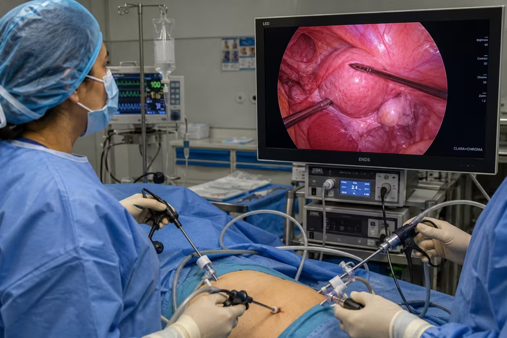
Diagnostic laparoscopy is the procedure that changes everything. A tiny high-definition camera — smaller than a pencil — enters the abdomen through a 1cm incision. Every structure in the pelvis is seen directly, in real time, under 10–20× magnification. What was invisible becomes visible. And because Dr. Pranay Shah performs diagnostic and operative laparoscopy in the same sitting, what is found is also fixed — without a second surgery, a second anaesthetic, or a second recovery.
Request a Free Callback
Why Normal Tests Don't Tell the Whole Story
This is the question that brings most patients to Dr. Shah’s surgical consultation: ‘My ultrasound is normal, my hormones are normal, my husband’s semen analysis is normal — so why am I not pregnant?’ The answer is almost always that there is a structural problem inside the pelvis that standard investigations simply cannot detect.
What Ultrasound Can See
- Large ovarian cysts (>2cm)
- Uterine fibroids that distort the uterine wall
- Hydrosalpinx (fluid-filled tube) when significantly enlarged
- Large endometrioma (chocolate cysts) of the ovary
- Uterine size and basic anatomy
- Dominant follicle growth and ovulation
- Good for: Hormonal monitoring, large structural abnormalities
What Ultrasound CANNOT See
- Peritoneal endometriosis – microscopic or fine implants on pelvic surfaces
- Pelvic adhesions – scar tissue between organs
- Mild or moderate endometriosis (Stage I and II)
- Tubal fimbrial damage or blockage at the far end
- Small submucosal fibroids (inside uterine cavity)
- The actual architecture of pelvic anatomy and organ mobility
- Cannot replace: Direct visual inspection of the pelvic cavity
How Often Does Laparoscopy Find Something Missed on Ultrasound?
Peritoneal endometriosis
Found in 30-50% of unexplained infertility cases at laparoscopy.
Pelvic adhesions
Found in 15-40% – often from prior infection, prior surgery, or ruptured appendix.
Tubal disease
Partial or complete blockage, fimbrial damage, or early hydrosalpinx formation in 10-30%.
Two Functions of One Procedure: Diagnostic + Operative
Laparoscopy at Wellspring is never just a “look inside.” Dr. Pranay Shah follows a fundamental principle: you do not put a patient under general anaesthesia for information alone. If something correctable is found, it is corrected in the same procedure – same anaesthesia, same incisions, same recovery. This is the single biggest advantage of operative laparoscopy over diagnostic-only scoping.
Diagnostic
- Endometriosis implants
- Adhesions / scar tissue
- Ovarian cysts
- Tubal patency (dye test)
- Fibroid location
- Pelvic anatomy assessment
- Chromopertubation result
Operative
- Endometriosis excision / ablation
- Adhesiolysis (cutting scar tissue)
- Cystectomy (cyst removal)
- Tubal cannulation / salpingectomy
- Laparoscopic myomectomy
- Ovarian drilling (PCOD)
- Peritoneal washing
Same Sitting
- One anaesthetic only
- One recovery period
- Lower total cost than two separate procedures
- Faster return to fertility attempts
- Reduced patient anxiety – no “wait and see”
- Treated tissue heals faster when managed immediately
When Dr. Pranay Shah Recommends Laparoscopy - and When He Does Not
Clinical honesty is the foundation of every surgical recommendation Dr. Shah makes. Laparoscopy is not a universal first-line investigation – it is a surgical procedure under general anaesthesia, and it is recommended only when the clinical picture justifies it.
| Timeframe | What You Experience | Activity Level |
|---|---|---|
| Day of procedure | Awake within 1-2 hours. Mild abdominal discomfort. Shoulder or upper abdominal pain (CO2 gas under the diaphragm) – common, resolves in 24-48 hours. Nausea from anaesthesia in some patients. | Resting. Accompanied discharge same evening for most patients. |
| Day 1-2 | Bloating and mild incision site tenderness. CO2 shoulder pain peaks and then resolves. Light activity at home. | Walking around the house. No driving. No heavy lifting. |
| Day 3-5 | Most patients feel significantly better. Incision soreness minimal – incisions are healing. Energy returning. | Light housework. Can sit comfortably and work from home. |
| Day 5-7 | Return to desk work / office for most patients. | Normal desk activity. Avoid heavy physical exertion. |
| Day 10-14 | Full physical recovery in the majority of cases for diagnostic laparoscopy. Operative cases with myomectomy or extensive adhesiolysis: 2-3 weeks. | Normal activity including exercise. |
| 3-6 months post-op | Uterine healing complete (myomectomy cases). Ovulation cycles re-established (ovarian drilling cases). Pelvic environment restored. | Active fertility attempts – natural, IUI, or IVF as planned with Dr. Shah. |
When to Contact Wellspring After Laparoscopy Serious complications after laparoscopy are rare – occurring in less than 1% of procedures in experienced hands. However, contact Dr. Shah’s team immediately if you experience: fever above 38°C (100.4°F) after the first 24 hours, increasing abdominal pain that is worsening not improving after Day 2, shoulder pain that is worsening after Day 3 (normal CO2 shoulder pain improves, not worsens), heavy vaginal bleeding beyond normal light spotting, difficulty passing urine or significant abdominal bloating, or any discharge or redness from the incision sites. Wellspring IVF post-surgery helpline: 9099946050.
Your Fertility Consultant
Our fertility specialists are committed to providing personalized, compassionate care with
the latest reproductive medicine techniques.
Dr. Pranay Shah
Divyesh Bhalodia
Urmi Chauhan
What Actually Happens During Laparoscopy - Step by Step
The word “surgery” understandably causes anxiety. Understanding exactly what happens – in plain language – transforms this from something frightening to something logical and manageable. Here is every step of a diagnostic and operative laparoscopy at Wellspring IVF:
Anaesthesia Administration
General anaesthesia is administered by a consultant anaesthetist. You are completely asleep and feel nothing throughout the procedure. The entire process is monitored with pulse oximetry, ECG, and blood pressure tracking.
Carbon Dioxide (CO2) Insufflation
A small needle (Veress needle) creates a tiny entry point at or near the navel. CO2 gas is gently introduced into the abdominal cavity. This creates space between the abdominal wall and the internal organs – giving Dr. Shah a clear, unobstructed field of view.
Trocar Placement - The "Ports"
2-3 small incisions (each 5-10mm – smaller than a centimetre) are made on the abdomen. Metal or plastic trocars (hollow tubes) are inserted through these incisions. These are the “ports” through which all instruments travel. There is no large abdominal cut.
Laparoscope Insertion & Diagnostic Survey
A slim laparoscope (a camera with fibre-optic light) is inserted through the central port. Dr. Shah performs a complete 360 degree systematic survey of all pelvic organs – uterus, both ovaries, both fallopian tubes, the pouch of Douglas (behind the uterus), the bowel surface, the bladder, and the peritoneum. This is the diagnostic phase. Everything is displayed in high definition on the operating theatre monitor.
Operative Treatment (Same Sitting)
If any pathology is found – endometriosis implants, fibroids, adhesions, blocked tubes, ovarian cysts, or PCOS follicles – Dr. Shah treats it immediately without a second procedure. Specialised instruments are inserted through the other ports. Energy devices (harmonic scalpel, bipolar diathermy) allow precise cutting, ablation, or coagulation with minimal blood loss.
Chromopertubation (Dye Test)
Blue methylene dye is injected through the uterine cervix during the procedure. Dr. Shah directly observes whether the dye flows freely out of each fallopian tube, spills freely into the pelvis, or is obstructed. This is the gold standard of tubal patency assessment – far more accurate than an X-ray HSG.
Instrument Removal & CO2 Release
All instruments are removed. The CO2 gas is released. The abdomen returns to normal. The tiny port sites are closed with 1-2 absorbable sutures or skin glue – no stitches to remove.
Recovery & Discharge
You wake up in the recovery room. Most patients are fully alert within 1-2 hours. At Wellspring, diagnostic laparoscopy is performed as a day-care procedure – the majority of patients are discharged the same day or with one overnight stay.
Dr. Pranay Shah on What Patients Fear Most: “The most common thing I hear is: ‘Doctor, I am scared of surgery.’ And my answer is always the same: what you are imagining is not what this is. You are imagining a large abdominal cut, weeks in hospital, and months of recovery. Laparoscopy is three incisions – each smaller than your fingernail. You arrive in the morning, the procedure takes 1 to 2 hours, and most of my patients go home the same evening. The fear of the procedure should never stand between you and the answer to why you are not conceiving.”
What Our Patients Say


A special thank you to Dr. Pranay Shah for his confidence, guidance, and the way he explained everything so patiently. His positive approach gave me so much strength, and today I am blessed with my baby.
Thank you to each and every member of the hospital for taking such great care of me and supporting me throughout this journey. Forever grateful. 💕

He is, without a doubt, the most patient doctor I have ever met. Of course, treatment can be done by many. What truly sets him apart is his maturity, the way he pauses, explains, comforts, and most importantly, seeks your permission before moving forward. You never feel rushed. You never feel unheard. You feel respected.
And the staff deserves equal appreciation. They handle even the most anxious and impatient moments with such calm grace and dignity that you slowly find your own heartbeat settling down. It feels less like a clinic and more like a safe space.
I wholeheartedly recommend him to anyone who overthinks, seeks reassurance, or simply needs a doctor who believes comfort is the first step of healing. With him, care begins long before the treatment does.

Highly recommend for their expertise, kindness and dedication. "Turned out dream into reality"



Our gratitude also extends to everyone in your clinic who offered a smile, reassurance, technical support, or a listening ear along the way. We feel incredibly blessed to have chosen your practice for our journey, and we will always cherish the precious gift you helped us receive.
Thank you, from the bottom of our hearts, for making our dream a reality.
Join 750+ Satisfied Families
Laparoscopy vs. Open Surgery - Why Keyhole Is the Standard of Care
For fertility patients, the choice between laparoscopic and open surgery is not simply a preference – it has direct consequences for recovery speed, adhesion formation, and how quickly you can attempt conception or IVF. Here is the complete comparison:
| Factor | Laparoscopy (Keyhole) | Open Surgery (Laparotomy) |
|---|---|---|
| Incision size | 3 small cuts – 5 to 10mm each (less than 1cm) | One large abdominal cut – 10 to 20cm |
| Scar visibility | Nearly invisible – fades within months | Prominent, permanent abdominal scar |
| Hospital stay | Day care or 1 night (majority of cases) | 3 to 5 days minimum |
| Return to normal activity | 5 to 7 days for desk work; 2 weeks full recovery | 4 to 6 weeks recovery |
| Post-operative pain | Mild – shoulder/upper abdomen discomfort (CO2 gas) for 1-2 days | Significant wound pain for 1-2 weeks |
| Blood loss | Minimal – controlled precision instruments | Higher – larger operative field |
| Infection risk | Very low – small entry points | Higher – larger wound surface |
| Pelvic adhesion risk post-op | Low – less tissue trauma, faster healing | Higher – large wound creates adhesion risk |
| Fertility recovery | Faster – conception attempts can begin within 1-2 cycles | Delayed – longer tissue healing required |
| Magnification | 10-20x magnification on HD monitor – sees what the naked eye cannot | Direct visual field – no magnification |
Why Adhesion Prevention Matters in Fertility Surgery Every surgery creates some degree of healing response. In the pelvis, aggressive tissue handling, large incisions, and prolonged exposure of raw surfaces can cause new scar tissue (adhesions) to form during healing – the very problem surgery was meant to address. Laparoscopy, by its minimally invasive nature, causes significantly less peritoneal trauma than open surgery. Combined with precise surgical technique – thorough irrigation, careful haemostasis, and anti-adhesion agents where appropriate – Dr. Shah minimises the risk of de novo adhesion formation after the procedure.
Conditions Diagnosed and Treated by Dr. Pranay Shah at Laparoscopy
Dr. Shah performs operative laparoscopy for the following fertility-affecting conditions. Each condition card explains what is found during the diagnostic survey and what is corrected during the operative phase of the same procedure.
Endometriosis
The most commonly missed cause of unexplained infertility – invisible on routine ultrasound in its early stages.
What Dr. Shah Finds at Laparoscopy
Endometriosis occurs when tissue similar to the uterine lining grows outside the uterus – on the ovaries, fallopian tubes, the back of the uterus (pouch of Douglas), the bladder, bowel surface, and the peritoneum. Mild endometriosis (Stage I and II) has no ultrasound signature whatsoever – it appears as microscopic deposits, fine powder-burn spots, or clear implants on the peritoneal surface. Dr. Shah identifies Stage I-IV endometriosis, including deep infiltrating endometriosis, retroversion of the uterus, and chocolate cysts (endometriomas) of the ovary.
What Is Treated in the Same Sitting
Excision of endometriotic implants from the peritoneum (preferred over ablation – removes the full lesion), endometrioma (chocolate cyst) cystectomy with maximum ovarian reserve preservation, lysis of peri-ovarian and peri-tubal adhesions, restoration of normal pelvic anatomy, and peritoneal washing to remove inflammatory fluid that impairs sperm function and embryo implantation.
Read more: Endometriosis — Detailed Guide
PCOD / PCOS – Laparoscopic Ovarian Drilling (LOD)
For medicine-resistant PCOD where clomiphene and gonadotropins have failed to trigger ovulation.
What Dr. Shah Finds at Laparoscopy
In PCOD, the ovaries contain multiple small follicles that fail to mature and rupture – causing chronic ovulation failure. Most patients with PCOD ovulate with oral medication or low-dose injectable gonadotropins. But in 15-20% of PCOD patients, medication resistance means months of failed cycles. Laparoscopy in this group allows Dr. Shah to directly assess both ovaries – their size, the density of follicles, the quality of the ovarian cortex – and confirm the PCOD morphology at direct visual inspection.
What Is Treated in the Same Sitting
Laparoscopic Ovarian Drilling (LOD): a diathermy needle or laser creates 4-8 small punctures (1-2mm deep) in the ovarian cortex of each ovary. Drilling reduces the androgen-producing stroma and corrects the LH:FSH hormonal imbalance – restoring a natural ovulatory cycle in 70-80% of previously anovulatory patients. It avoids OHSS and multiple pregnancy risk associated with gonadotropins.
Read more: PCOD/PCOS — Complete Guide
Uterine Fibroids – Laparoscopic Myomectomy
Surgical removal of fibroids that distort the uterine cavity or impair implantation.
What Dr. Shah Finds at Laparoscopy
Not all fibroids impair fertility. The critical distinction is location. Submucosal fibroids (inside the cavity) and large intramural fibroids (within the uterine wall, >4-5cm) directly impair implantation and increase miscarriage risk. Subserosal fibroids (on the outer surface) rarely affect fertility unless very large. Laparoscopy allows Dr. Shah to assess all fibroids – their exact position relative to the endometrial cavity, vascularity, and number – with a level of detail unavailable on ultrasound alone.
What Is Treated in the Same Sitting:
- Laparoscopic myomectomy: surgical removal of fibroids through keyhole incisions, preserving the uterus entirely
- The fibroid is enucleated from the uterine wall and extracted through the ports — uterus is repaired in layers with absorbable sutures under magnification
- Unlike open myomectomy: less blood loss, less adhesion formation, faster healing, earlier return to fertility
- Note: Very large fibroids (>10–12cm) or multiple fibroids (>5) may require open myomectomy — Dr. Shah advises honestly on the appropriate approach for each patient
- Post-myomectomy waiting: 3–6 months before IVF or conception attempts to allow complete uterine healing
→ Read more: Uterine Fibroids — Detailed Guide
Blocked Fallopian Tubes, Hydrosalpinx & Tubal Disease
Including the mandatory salpingectomy before IVF for hydrosalpinx patients.
What Dr. Shah Finds at Laparoscopy
Fallopian tube disease is assessed during the diagnostic phase using chromopertubation – blue dye injected through the cervix while Dr. Shah directly watches both tubes under the laparoscope. A normal tube shows free spill of dye from the fimbriated end into the pelvis. A blocked tube shows no spill. A hydrosalpinx (fluid-filled tube) is visible as a swollen, sausage-shaped structure – and is a fertility emergency because the toxic fluid inside leaks back into the uterus and actively destroys embryos during IVF.
What Is Treated in the Same Sitting:
- Tubal cannulation: for proximal (near the uterus) blockages, a fine wire is passed through the tube under laparoscopic guidance to open the blockage
- Fimbrioplasty / fimbriolysis: opening of minor fimbrial adhesions at the far end of the tube
- Salpingectomy (tube removal) for hydrosalpinx: MANDATORY before IVF. The toxic fluid in a hydrosalpinx reduces IVF success rates by 50%. Removing the damaged tube before embryo transfer restores the uterine environment and significantly improves IVF outcomes
- Adhesiolysis around tubes: freeing tubes trapped by endometriosis or post-infection scar tissue
→ Read more: Blocked Fallopian Tubes — Complete Guide
Pelvic Adhesions – Adhesiolysis
Scar tissue that traps organs, distorts tubal anatomy, and blocks the path of sperm to egg.
What Dr. Shah Finds at Laparoscopy
Pelvic adhesions are bands of scar tissue that form between organs – ovary to tube, tube to uterus, uterus to bowel, ovary to pelvic wall. They are completely invisible on ultrasound. Their causes include prior pelvic infection (PID), prior abdominal or pelvic surgery, ruptured appendix, prior laparoscopy with poor technique, and severe endometriosis. Adhesions fix organs in abnormal positions, prevent the fallopian tube from picking up the egg after ovulation, and can kink or occlude the tube entirely.
What Is Treated in the Same Sitting:
- Adhesiolysis: using scissors, harmonic scalpel, or laser energy, Dr. Shah systematically cuts through adhesion bands under direct magnified vision
- Organ mobility is restored — the ovary, tube, and uterus return to their natural anatomical positions
- Fine peritoneal adhesions overlying the tube are removed to restore the tube’s natural ‘sweeping’ function
- Anti-adhesion agents (Interceed, Seprafilm) may be applied to raw surfaces to reduce the likelihood of adhesion reformation during healing
- Post-adhesiolysis: natural conception attempts in the first 3–6 months, when the pelvis is at its most normal state, offer the best
→ Read more: Blocked Fallopian Tubes — Adhesion Section
Talk to Dr. Shah About Cavity Optimisation
When Dr. Pranay Shah Recommends Laparoscopy - and When He Does Not
Clinical honesty is the foundation of every surgical recommendation Dr. Shah makes. Laparoscopy is not a universal first-line investigation – it is a surgical procedure under general anaesthesia, and it is recommended only when the clinical picture justifies it.
Laparoscopy IS Recommended When:
- Unexplained infertility after 12 months of attempting (or 6 months if age >35) with normal basic investigations
- Suspected endometriosis – chronic pelvic pain, painful periods, pain with intercourse, or elevated CA-125
- Confirmed or suspected tubal blockage on HSG (X-ray dye test) requiring direct visual confirmation and possible treatment
- Hydrosalpinx on ultrasound or HSG – MANDATORY removal before IVF
- PCOD with confirmed anovulation despite adequate medical treatment (clomiphene resistance)
- Fibroid found on ultrasound close to the uterine cavity – to assess true impact before IVF decision
- Prior pelvic infection (PID) or prior abdominal surgery – high clinical suspicion of adhesions
- Two or more failed IVF cycles elsewhere with otherwise good embryos – to rule out correctable pelvic pathology
- Prior ectopic pregnancy – to assess the remaining tube and pelvic anatomy
Laparoscopy Is NOT Recommended When:
- Young couple (<35), less than 12 months of trying, no symptoms, normal ultrasound and hormones – watchful expectancy is appropriate first
- Male factor infertility is the sole identified cause – surgery on the female partner does not address a sperm problem
- When IVF is already the planned path (not all blocked tubes or mild endometriosis need surgical correction before IVF – Dr. Shah evaluates case by case)
- Active pelvic infection – laparoscopy during active infection carries higher complication risk
- Severe anaesthesia risk from medical comorbidities – clinical risk-benefit assessment required
Dr. Shah’s guiding principle: “I will not perform surgery for a problem I am not certain exists, and I will not withhold surgery for a problem that is preventing pregnancy. Every recommendation is explained with clinical reasoning – not protocol.”
Laparoscopy and IVF - How Surgery Maximises Your IVF Success
| Clinical Situation | Without Laparoscopy First | With Laparoscopy First | Dr. Shah’s Recommendation |
|---|---|---|---|
| Hydrosalpinx present | IVF success rate reduced by ~50% – toxic fluid contaminates uterine environment at embryo transfer | Salpingectomy removes the toxic source. IVF success restored to expected rates for age/embryo quality | Laparoscopy + salpingectomy MANDATORY before IVF. Non-negotiable. |
| Stage III-IV Endometriosis | Heavy peritoneal inflammation, ovarian damage, distorted anatomy – all impair egg quality and implantation | Surgical clearance improves egg retrieval, reduces inflammation, restores anatomy | Strongly recommended before IVF – particularly for large endometriomas |
| Stage I-II (Mild) Endometriosis | Moderate evidence for benefit of surgical treatment before IVF – clinical debate exists | Excision removes inflammatory load and may improve implantation | Case-by-case – depends on patient age, embryo reserve, and prior IVF history |
| Submucous fibroid distorting cavity | Fibroid directly competes with embryo for implantation space – failed transfer likely | Myomectomy removes the distortion. Uterine cavity normalised before embryo transfer | Recommended – hysteroscopic or laparoscopic depending on fibroid type and size |
| Pelvic adhesions suspected | Tubes may be blocked/kinked – failed egg pickup. Anti-adhesion environment hostile to IVF | Adhesiolysis frees organs, restores normal pelvic environment | Recommended when suspicion is high – particularly post-infection or post-surgical |
| Normal anatomy, first IVF attempt | Proceeding directly to IVF is standard | No benefit from exploratory laparoscopy before first attempt with no clinical indication | NOT recommended – first IVF attempt with no findings should proceed without surgery |
After Laparoscopy — What Comes Next at Wellspring
Surgery is not the destination — it is the preparation. After laparoscopy, Dr. Shah maps the next clinical step based on what was found and corrected:
IVF / ICSI Treatment
For patients with bilateral tube disease, older patients (>35), low ovarian reserve, or male factor alongside corrected pelvic pathology. Read the IVF Treatment Hub.
IUI Treatment
For patients with patent tubes, good ovarian reserve, and mild male factor – IUI immediately post-surgery has high natural conception rates. Read more for IUI treatment Guide.
Natural conception window
For patients under 35 with single-factor disease (mild endometriosis, adhesiolysis, PCOD drilling): 3-6 months of natural attempts before ART is recommended.
Hysteroscopy
Frequently combined with laparoscopy on the same day to assess the uterine cavity simultaneously – one anaesthetic, complete pelvic + uterine evaluation.
Recovery After Laparoscopy — What to Expect
One of the biggest reasons patients delay necessary laparoscopy is fear of a long, painful recovery. The reality of keyhole surgery recovery is significantly gentler than patients anticipate.
Timeframe | What You Experience | Activity Level |
|---|---|---|
Day of procedure | Awake within 1–2 hours. Mild abdominal discomfort. Shoulder or upper abdominal pain (CO₂ gas under the diaphragm) — common, resolves in 24–48 hours. Nausea from anaesthesia in some patients. | Resting. Accompanied discharge same evening for most patients. |
Day 1–2 | Bloating and mild incision site tenderness. CO₂ shoulder pain peaks and then resolves. Light activity at home. | Walking around the house. No driving. No heavy lifting. |
Day 3–5 | Most patients feel significantly better. Incision soreness minimal — incisions are healing. Energy returning. | Light housework. Can sit comfortably and work from home. |
Day 5–7 | Return to desk work / office for most patients. | Normal desk activity. Avoid heavy physical exertion. |
Day 10–14 | Full physical recovery in the majority of cases for diagnostic laparoscopy. Operative cases with myomectomy or extensive adhesiolysis: 2–3 weeks. | Normal activity including exercise. |
3–6 months post-op | Uterine healing complete (myomectomy cases). Ovulation cycles re-established (ovarian drilling cases). Pelvic environment restored. | Active fertility attempts — natural, IUI, or IVF as planned with Dr. Shah. |
When to Contact Wellspring After Laparoscopy
Serious complications after laparoscopy are rare — occurring in less than 1% of procedures in experienced hands. However, contact Dr. Shah’s team immediately if you experience:
- Fever above 38°C (100.4°F) after the first 24 hours
- Increasing abdominal pain that is worsening, not improving, after Day 2
- Shoulder pain that is worsening after Day 3 (normal CO₂ shoulder pain improves, not worsens)
- Heavy vaginal bleeding beyond normal light spotting
- Difficulty passing urine or significant abdominal bloating
- Any discharge or redness from the incision sites
📞 Wellspring IVF post-surgery helpline: 9099946050 — available for all post-operative concerns.
Frequently Asked Questions
Is laparoscopy painful? I am very anxious about surgery.
Laparoscopy is performed under general anaesthesia – you are completely asleep and feel nothing during the procedure. After surgery, the most common discomfort is not from the incisions (which are tiny) but from residual CO2 gas under the diaphragm causing referred pain to the shoulder and upper abdomen. This is a pressure sensation, not a wound pain – it resolves completely within 24-48 hours as the gas is absorbed. Most patients rate their post-laparoscopy pain as 2-4 out of 10 and manage it well with standard painkiller tablets. The anxiety before the procedure is almost always greater than the discomfort after it.
What is the difference between diagnostic laparoscopy and operative laparoscopy?
Diagnostic laparoscopy means inserting the camera to assess and identify any pathology. Operative laparoscopy means the surgeon also treats the identified pathology during the same procedure using additional instruments through the other ports. At Wellspring, Dr. Shah always performs both functions together – you are not woken up with a “we found something, we need to schedule a second procedure” outcome. If it is findable and fixable, it is fixed in the same sitting under the same anaesthesia.
How long does the laparoscopy take?
A pure diagnostic laparoscopy (look and dye test only) takes approximately 30-45 minutes from first incision to skin closure. When operative procedures are performed, the total time depends on the findings: endometriosis excision adds 30-60 minutes, ovarian drilling 15-20 minutes, adhesiolysis 30-60 minutes depending on severity, and laparoscopic myomectomy 60-120 minutes for moderate-sized fibroids. Dr. Shah discusses the anticipated duration with you during your pre-operative consultation based on your specific findings and planned procedures.
Will laparoscopy affect my ovarian reserve or egg count?
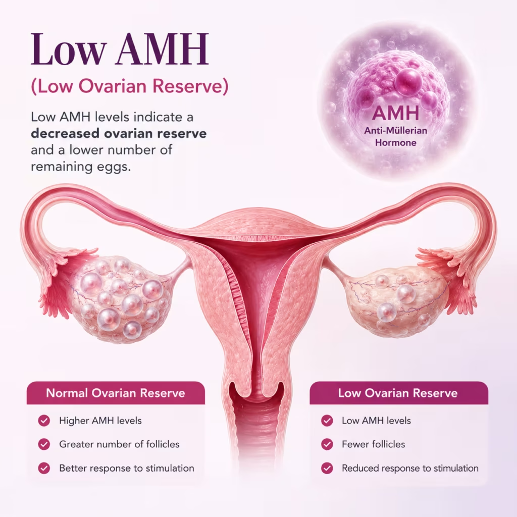
Standard diagnostic laparoscopy does not affect ovarian reserve. Ovarian drilling for PCOD causes a modest, temporary reduction in AMH (anti-Mullerian hormone) in some patients – this typically recovers within 3-6 months. The most important consideration is endometrioma cystectomy: removing a chocolate cyst unavoidably removes some healthy ovarian tissue along with the cyst wall. Dr. Shah uses the “stripping technique” to minimise normal ovarian tissue loss during endometrioma removal. For patients with already-reduced ovarian reserve (low AMH), the risk-benefit of endometrioma surgery versus proceeding directly to IVF is discussed in detail before any surgical recommendation is made.
Do I need to be admitted overnight?
For standard diagnostic laparoscopy with or without minor operative procedures (ovarian drilling, mild adhesiolysis, tubal dye test), the procedure is performed as a day-care case at Wellspring – you arrive in the morning, the procedure is completed, and you are discharged the same evening with an accompanying person. For more extensive operative procedures – laparoscopic myomectomy, severe endometriosis excision, or significant adhesiolysis – one overnight stay may be recommended for monitoring. Dr. Shah discusses expected hospital stay during your pre-operative consultation.
My previous doctor said I have a hydrosalpinx. Is surgery really necessary before IVF?
Yes – absolutely, and this is non-negotiable based on current evidence. A systematic review of published studies (Cochrane, ESHRE) shows that the presence of a hydrosalpinx reduces IVF live birth rates by approximately 50% compared to patients without hydrosalpinx. The mechanism is well-established: the toxic fluid inside a hydrosalpinx periodically refluxes into the uterine cavity, creating an embryo-hostile environment at the time of transfer. Salpingectomy (surgical removal of the damaged tube) before IVF consistently doubles live birth rates in affected patients. This is not a commercial surgical recommendation – it is a clinical evidence-based requirement. The tube removed was already non-functional for natural conception. Read more on Blocked Fallopian Tubes.
What is the diagnostic laparoscopy cost in India, and specifically at Wellspring Ahmedabad?
While the average diagnostic laparoscopy cost in India varies across different tiers of cities, at Wellspring IVF, we ensure our pricing is completely transparent and value-driven. The overall cost depends on whether the procedure remains purely diagnostic or transitions into an operative intervention (such as treating endometriosis or clearing adhesions).
Dr. Shah provides a complete, itemised cost estimate during your pre-operative consultation, ensuring there are no hidden fees. For current packages and customized pricing, please reach out directly to our team at 9099946050.
Related Insights & Articles
 Blocked fallopian tubes can make it hard for a woman to get pregnant. But there are ways to treat this…June 11, 2026
- Are you feeling worried about low AMH and IVF? You are not alone. Many women have to face the same…June 11, 2026
- Can You Increase AMH Levels Naturally? What Science Says About Low AMH and Fertility Quick Answer In most cases, AMH…June 11, 2026